LAB 2 : MEASUREMENT & COUNTING OF CELLS USING
MICROSCOPE
INTRODUCTION
An ocular micrometer is a glass disk that fits in a microscope eyepiece that has a ruled scale, which is used to measure
the size of magnified objects. The physical length of the marks on the scale
depends on the degree of magnification. The ruler on a typical ocular
micrometer has between 50 to 100 individual marks, is 2 mm long and has a distance
of 0.01 mm between marks.
The procedure to use ocular micrometer is
firstly, measure
the actual size of the letter on the microscope slide using the millimeter
ruler. This measurement will help you calibrate the ocular micrometer to
determine if it is giving you accurate measurements.
Then, attach the ocular micrometer to the microscope
eyepiece by unscrewing the eyepiece cap, placing the ocular micrometer over the
lens and screwing the eyepiece cap back into place. Some microscopes may have
an ocular micrometer pre-installed, allowing you to skip this step.
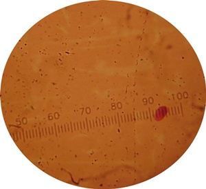
Figure
: ocular scale (above) and stage micrometer scale (below)
The
procedure in using Neubauer Chamber
is firstly, Ensure that the special cover slip provided with the counting
chamber (thicker than standard cover slips and with a certified flatness) is
properly positioned on the surface of the counting chamber. When the two glass
surfaces are in proper contact Newton's
rings can be observed. If so, the cell suspension is applied to
the edge of the cover slip to be sucked into the void by capillary
action which completely fills the chamber with the sample.
Looking at the chamber through a microscope,
the number of cells in the chamber can be determined by counting. Different
kinds of cells can be counted separately as long as they are visually
distinguishable. The number of cells in the chamber is used to calculate
the concentration or density of the cells in
the mixture the
sample comes from. It is the number of cells in the chamber divided by the
chamber's volume (the chamber's volume is known from the start).
RESULT
2.1
Ocular Micrometer
Yeast
400x magnification :
5 X 0.0025mm = 0.0125mm
figure : yeast 400x magnification
1000x magnification :
7 x 0.0011mm = 0.0077mm
figure : yeast 1000x magnification
Lactobacillus sp.
400x magnification :
2 x 0.0025mm = 0.005mm
figure : Lactobacillus sp. 400x magnification
1000x magnification :
5 x 0.0011mm = 0.0055mm
figure : Lactobacillus sp. 1000x magnification
2.2
Neubauer Chamber
sum of the
cell 10 box = 642
average =
642/10 = 64.2
volume box
(16 box) = 0.2 mm x 0.2 mmx 0.1 mm
= 4 x 10^-3 mm
= 4 x 10^-6 cm^3
64.2 cell in
4 x 10 ^-6 ml
1 ml =
642/ 4 x 10 ^-6 = 1.61 x 10^8 cell/ml
DISCUSSION
2.1
Ocular Micrometer
magnification X400.
5 X 0.01 = 0.05
0.05/20 = 2.5 micrometer
0.05/20 = 2.5 micrometer
Suppose the full length of the reticle scale covered 25
divisions of the stage micrometer.
Then the full length of the reticle scale is equivalent to
(25 x 0.1mm) = 2.5mm long.
For an eyepiece reticle with 100 divisions, each division
will measure 25µm at the stage for this
magnification.
magnification X1000.
5 X 0.01 = 0.05
0.05/47 = 1 micrometer
Selecting
the X100 objective and repeating the exercise above would show that the reticle
scale
now covers 10 divisions of the stage scale.
Then the full length of the reticle scale is equivalent to
(10 x 0.1mm) = 1mm long.
For an eyepiece reticle with 100 divisions, each division
will measure 10µm at the stage for this
magnification.
In summary you can apply these conversion factors to state
what each division of the eyepiece
reticle is measuring for a selected magnification.
X400 : 1 division = 25µm
X1000 : 1 division =
10µm
2.2
Neubauer Chamber
Suppose that we
conduct a count on 10 box, and count 642 particles in all 10 small squares described. Each square has an
area of 0.04 mm-squared and depth of 0.1 mm. The total volume in each square is
(0.04)x(0.1) = 0.004 mm-cubed. We have
10 squares with combined volume of 10x(0.004) = 0.04 mm-cubed. Thus we counted 642 particles in a volume of 0.004
mm-cubed, giving you 642/0.04 = 1.6 x 10^4 particles per mm-cubed. There are
1000 cubic millimeters in one cubic centimeter (same as a milliliter), so your
particle count is 16,050,000 per ml.
CONCLUSION
Ocular micrometers have no units on them - they are like a
ruler with marks but no numbers. In order to use one to measure something under
a microscope, you must assign numbers to the marks. This is done by looking
through your ocular micrometer at a stage micrometer mounted on a slide. The
stage micrometer is just a ruler with fixed known distances, so you can use it
to tell how far apart marks are on the ocular micrometer.
This has to be done because the marks on the ocular
micrometer are different distances apart depending on the magnification used on
the microscope. It must be calibrated for each objective.
Cell culture and many applications that require use of
suspensions of cells it is necessary to determine cell concentration. One can
often determine cell density of a suspension spectrophotometrically, however
that form of determination does not allow an assessment of cell viability, nor
can one distinguish cell types.
http://en.wikipedia.org/wiki/Ocular_micrometer
academic.evergreen.edu/curricular/fcb/wk2calibration.doc
academic.evergreen.edu/curricular/fcb/wk2calibration.doc
en.wikipedia.org/wiki/Hemocytometer
sfiles.crg.es/protocols/cellculture/img/neubauer.jpg/view


No comments:
Post a Comment