LAB 2: MEASUREMENT AND COUNTING OF CELLS USING MICROSCOPE by NUR DIANA BT ABDUL JALIL (114120)

2.1 Ocular Micrometer

INTRODUCTION

Ocular Microscope

Microscopes are often used to measure small objects. For instance, forensic scientists use microscopy to measure the distance between microscopic tool marks or the diameter of fibers. Compound microscopes have ocular micrometers, or rulers that can help you measure items under the microscope. However, the scale on the ocular micrometer changes with total magnification, and thus has no absolute value. Therefore, the ocular micrometer does not have units and it needs to be calibrated prior to use. 

We use a stage micrometer to calibrate the ocular micrometer. A stage micrometer is essentially a ruler that is mounted on a microscope slide that does have units (millimeters (mm) or micrometers (mm)). When calibrating, we will line up the stage micrometer with the ocular micrometer and count the number of divisions on the ocular micrometer per millimeter or micrometer on the staged micrometer. The number of divisions will change as the magnification changes.


OBJECTIVE

To measure and count cells using a microscope

Results

l.Lactobacillus sp.
                                                                  400x magnification

                                                                  1000x magnification

2. Yeast
                                                                400x magnification

                                                                 1000x magnification

calibration:

400x: 
5 x 0.01 = 0.05
0.05/ 20 = 0.0025 mm

1000x:
5 x 0.01 = 0.05mm
0.05/ 47 = 0.0011 mm

·         Lactobacillus sp.
    400x magnification : 
     2 division x 0.0025 mm = 0.005 mm
    
     1000x magnification : 
      5 division x 0.0011 mm = 0.0055 mm

·         Yeast
      400x magnfication :
      5 division x 0.0025 mm = 0.0125 mm
       
      1000x magnification :
       7 division x 0.0011 mm = 0.0077 mm


DISCUSSION

An ocular micrometer is a small glass disk with thin lines and numbering etched in the glass.
An ocular micrometer was placed into one ocular on your microscope so that the lines superimposed on the image will allow to measure the specimen.
For each magnification, must compare the lines on the ocular micrometer to the lines on a stage micrometer.
The stage micrometer is a glass slide with precisely space lines etched at known intervals.
The vertical distance of an object that is in focus.  When magnification is increased, less of the object is in focus (depth of field decreases) – but greater detail of the area in focus can be seen.
We can adjust the focus of our eyepiece in order to make the scale as sharp as possible. If we do that, also adjust the other eyepiece to match the focus. Any ocular scale must be calibrated, using a device called a stage micrometer
The ocular lenses usually magnify 10X.  Thus the total magnification observed is the multiplication of the power of magnification of the ocular times the objective.  For example an object magnified by the ocular and the 40X high-dry objective is viewed at 4002 times its real size.  Most ocular lenses can be moved back and forth to adjust to the interpupillary distance of the student.  When first using the microscope, adjust the ocular lenses back and forth until a circular field is viewed with both eyes open.  Additionally, many microscopes allow the ocular lenses to be adjusted up and down (mechanical tube length adjustment) and there is a scale alongside the tube.  After adjusting the interpupillary distance, read the distance off the scale and adjust the tube length of the ocular lens to the same value.  Now the ocular lenses are adjusted to our eyes.

Conclusion
Ocular micrometer a glass disk that fits in a microscope eyepiece and that has a ruled scale when calibrated with a slide micrometer, direct measurements size of a microscopic object can be made.

Reference


2.2 Neubauer Chamber


INTRODUCTION

Neubauer Chamber

Neubauer chamber or hemocytometer are more convenient for counting microbes. It is a heavy glass slide with two counting areas separated by a H-shaped through figure. To prepare the counting chamber the mirror-like polished surface is carefully cleaned with lens paper. The coverslip is also cleaned. Coverslips for counting chambers are specially made and are thicker than those for conventional microscopy, since they must be heavy enough to overcome the surface tension of a drop of liquid. The coverslip is placed over the counting surface prior to putting on the cell suspension. The suspension is introduced into one of the V-shaped wells with a pasteur or other type of pipet. The area under the coverslip fills by capillary action. Enough liquid should be introduced so that the mirrored surface is just covered. The charged counting chamber is then placed on the microscope stage and the counting grid is brought into focus at low power.

It is essential to be extremely careful with higher power objectives, since the counting chamber is much thicker than a conventional slide. The chamber or an objective lens may be damaged if the user is not not careful. One entire grid on standard hemacytometers with Neubauer rulings can be seen at 40x (4x objective). The main divisions separate the grid into 9 large squares (like a tic-tac-toe grid). Each square has a surface area of one square mm, and the depth of the chamber is 0.1 mm. Thus the entire counting grid lies under a volume of 0.9 mm-cubed.


Objective
1.      To count cells using microscope.

RESULT


sum of the cell 10 box = 642
average = 642/10 = 64.2

volume box (16 box) = 0.2 mm x 0.2 mmx 0.1 mm
                                 = 4 x 10^-3 mm
                                 = 4 x 10^-6 cm^3
64.2 cell in 4 x 10 ^-6 ml
1 ml = 642/ 4 x 10 ^-6 = 1.61 x 10^8 cell/ml

  
DISCUSSION
To prepare the counting chamber the mirror-like polished surface is carefully cleaned with lens paper. The coverslip is also cleaned.
Coverslips for counting chambers are specially made and are thicker than those for conventional microscopy, since they must be heavy enough to overcome the surface tension of a drop of liquid.
The coverslip is placed over the counting surface prior to putting on the cell suspension. The suspension is introduced into one of the H-shaped wells with a pasteur or other type of pipet.
The area under the coverslip fills by capillary action. Enough liquid should be introduced so that the mirrored surface is just covered.
The charged counting chamber is then placed on the microscope stage and the counting grid is brought into focus at low power.


Conclusion
Neubauer chamber is a device used for determining the number of cells per unit volume of a suspension. Hemocytometer was widely used since it was originally designed for performing blood cell counts.


Reference


LAB 3 : ZAYANI BINTI MUKHTAR

LAB 3 : PREPARATION AND STERILIZATION OF CULTURE  MEDIA


INTRODUCTION


growth medium or culture medium is a liquid or gel designed to support the growth of microorganisms or cells, or small plants. There were different types of culture media for different types of cell growing. It is depends on the culture in the experiment used. There are two major types of growth media: those used for cell culture, which use specific cell types derived from plants or animals, and microbiological culture, which are used for growing microorganisms, such as bacteria or yeast. The most common growth media for microorganisms are nutrient broths and agar plates; specialized media are sometimes required for microorganism and cell culture growth. In this experiment both agar and the nutrient broth are been prepared. 


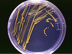
An agar plate -- an example of a bacterial growth medium.


Autoclaving is one of the techniques in the heat sterilization. An autoclave is an instrument used to sterilize equipment and supplies by subjecting them to high pressure saturated steam at 121 °C for around 15–20 minutes depending on the size of the load and the contents. Autoclaving uses steam under high pressure. Autoclaving is the most effective and most efficient means of sterilization.


Autoclave in laboratory


DISCUSSION


In this experiment we need to prepare our own culture media. The most common growth media for microorganisms are nutrient broths (liquid nutrient medium) or LB medium (Lysogeny Broth). Liquid media are often mixed with agar and poured into Petri dishes to solidify. These agar plates provide a solid medium on which microbes may be cultured. They remain solid, as very few bacteria are able to decompose agar. Bacteria grown in liquid cultures often form colloidal suspensions . For our broth in this experiment we used :

0.6 g/L "Lab- lemco" powder (a beef extract)
0.4 g/L yeast extract
1.0 g/L peptone (a nitrogen source)
1.0 g/L sodium chloride 
3.0 g/L agar powder


Nutrient agar is a microbiological growth medium commonly used for the routine cultivation of non-fastidious bacteria. It is useful because it remains solid even at relatively high temperatures. Also, bacteria grown in nutrient agar grows on the surface, and is clearly visible as small colonies. In nutrient broth, the bacteria grows in the liquid, and is seen as a soupy substance, not as clearly distinguishable clumps. The distilled water is used for pH adjustment. Peptones are derived from animal milk or meat digested by proteolytic digestion. In addition to containing small peptides, the resulting spray-dried material includes fats, metals, salts, vitamins and many other biological compounds. Peptone is used in nutrient media for growing bacteria and fungi. 


 The preparation of the nutrient agar.

Autoclaves use pressurized steam to destroy microorganisms, and are the most dependable systems available for the decontamination of laboratory waste and the sterilization of laboratory glassware, media, and reagents. Higher temperature in autoclaving ensure that more rapid killing for the microorganisms. In autoclave, for efficient heat transfer, steam must flush the air out of the autoclave chamber. Before using the autoclave, check the drain screen at the bottom of the chamber and clean if blocked. If the sieve is blocked with debris, a layer of air may form at the bottom of the autoclave, preventing efficient operation. For efficient heat transfer, steam must flush the air out of the autoclave chamber. If the drain screen is blocked with debris, a layer of air may form at the bottom of the autoclave and prevent proper operation. The most important things is to make sure that the door is sealed before selecting the cycle.

Nutrient agar that will undergoing autoclaving.

CONCLUSION
In this experiment we had determined the correct ways to prepare a culture media use in microorganisms growing. We had weight the substances in the nutrient agar. During the experiment, we had taken few steps of sterilization to avoid contamination for our growth media.


REFERENCES


LAB 3 : PREPARATION AND STERILIZATION OF CULTURE MEDIA by Nurul Samihah binti Mohd Jamil (111408)

Introduction:


A growth medium or culture medium is a liquid or gel designed to support the growth of microorganisms or cells, or small plants like the moss Physcomitrella patens. There are different types of media for growing different types of cells.
An agar plate
There are two major types of growth media: those used for cell culture, which use specific cell types derived from plants or animals, and microbiological culture, which are used for growing microorganisms, such as bacteria or yeast. The most common growth media for microorganisms are nutrient broths and agar plates; specialized media are sometimes required for microorganism and cell culture growth. Some organisms, termed fastidious organisms, require specialized environments due to complex nutritional requirements. Viruses, for example, are obligate intracellular parasites and require a growth medium containing living cells.

Objectives: 
  • To prepare sterile nutrient agar for culturing microorganisms.
 Discussion:

To prepare agar; weigh 7.00 g of nutrient powder by using analytical balance and put in the scott bottle. Measure 250 ml of distilled water by measuring cylinder and add up in the scott bottle containing nutrient powder. Then, mixed up nutrient media with distilled water. Recap the bottles loosely and set aside for the sterilization. Repeat the  steps for 10.45g in 100 ml MRS broth and 5.00 g in 250 ml peptone 2%. After that, sterilize  all media at 121˚C for 15 minutes by using autoclave.


A widely-used method for heat sterilization is the autoclave, sometimes called a converter. Autoclaves commonly use steam heated to 121–134 °C (250–273 °F). To achieve sterility, a holding time of at least 15 minutes at 121 °C (250 °F) or 3 minutes at 134 °C (273 °F) is required. Additional sterilizing time is usually required for liquids and instruments packed in layers of cloth, as they may take longer to reach the required temperature (unnecessary in machines that grind the contents prior to sterilization). Following sterilization, liquids in a pressurized autoclave must be cooled slowly to avoid boiling over when the pressure is released. Modern converters operate around this problem by gradually depressing the sterilization chamber and allowing liquids to evaporate under a negative pressure, while cooling the contents. Proper autoclave treatment will inactivate all fungi, bacteria, viruses and also bacterial spores, which can be quite resistant. It will not necessarily eliminate all prions.


Autoclaves are widely used in microbiology, medicine, tattooing, body piercing, veterinary science, mycology, dentistry, chiropody and prosthetics fabrication. They vary in size and function depending on the media to be sterilized.
Typical loads include laboratory glassware, surgical instruments, medical waste, patient pair utensils, animal cage bedding, and lysogeny broth.
A notable growing application of autoclaves is the pre-disposal treatment and sterilization of waste material, such as pathogenic hospital waste. Machines in this category largely operate under the same principles as conventional autoclaves in that they are able to neutralize potentially infectious agents by utilizing pressurized steam and superheated water. A new generation of waste converters is capable of achieving the same effect without a pressure vessel to sterilize culture media, rubber material, gowns, dressing, gloves, etc. It is particularly useful for materials which cannot withstand the higher temperature of a hot air oven. For all-glass syringes, sterilizing in a hot air oven is a better method.
Autoclaves are also widely used to cure composites and in the vulcanization of rubber. The high heat and pressure that autoclaves allow help to ensure that the best possible physical properties are repeatably attainable. The aerospace industry and sparmakers (for sailboats in particular) have autoclaves well over 50 feet long, some over 10 feet wide.

Conclusion:
 
This report was identified the correct way to prepare a culture media. The type of culture media was used nutrient agar which prepare suitable medium for microorganisms growth. To culture the microorganisms in the nutrient agar, few steps of sterilization was taken to avoid any contamination on the colony. Autoclaving is the process used to sterilize the nutrient agar. The media was inserted into an autoclave which is a large pressure cooker. The chamber provided high temperature and pressurized steam
References: 

ZASS's Guests

Bioprocess Technology ? ? ?

Bioprocess technology is the industrial application of biological processes involving living cells or their components to effect desired transformation of substrates. The major advantages of bioprocesses . . .

I'm interested to . . .

ZASS Technologists Bhd.

WELCOME to ZASS Technologists Bhd.

Bioprocess Technologist 1

Bioprocess Technologist 1
Zayani bt Mukhtar

Bioprocess Technologist 2

Bioprocess Technologist 2
Nur Diana bt Abdul Jalil

Bioprocess Technologist 3

Bioprocess Technologist 3
Nor Shaqira bt Azlan

Bioprocess Technologist 4

Bioprocess Technologist 4
Nurul Samihah binti Mohd Jamil

Interesting ? Let join us now !

Powered by Blogger.